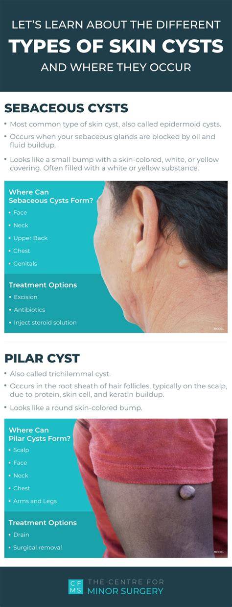
Epidermal inclusion cysts, often misnamed sebaceous cysts, are benign, slow-growing skin lesions that most commonly form under the surface of the skin and originate from the infundibulum or upper portion of a hair follicle. They can appear anywhere on the body but are frequently seen on the face, scalp, chest, back, and trunk.
What does an epidermal inclusion cyst look like?
A cyst is typically a round, dome-shaped bump located just under the skin surface. It may appear yellow or pale, and skin-colored in tone. Some cysts have a punctum-a dark dot in the center connecting to the skin surface. They can range from about 0.25 inches to more than 2 inches in diameter and may grow slowly over time. Skin discoloration is possible, often pink to red or darker than the surrounding skin, and a cyst can be tender or feel warm to the touch. In some cases, the cyst can move slightly under the skin.
What are epidermal inclusion cysts filled with?
Epidermal inclusion cysts contain keratin and cell debris. When drained by a dermatologist, the material appears thick and yellow and may have a foul odor.
Are epidermal inclusion cysts painful?
EICs are not normally painful. They can become inflamed or tender when touched, and pain may occur if the cyst grows or ruptures. Itching is possible at the site. Seek medical advice if pain develops or other concerning symptoms appear.
Where do epidermal inclusion cysts form?
EICs can form anywhere on the body but are most common on the face, chest, back, scalp, neck, legs, arms, and genitalia.
What causes an epidermal inclusion cyst?
EICs form after a blockage at the hair follicle’s infundibulum, where skin cells are shed as they life cycle ends. Injury to the skin, such as a scratch, surgical wound, acne, or chronic sun damage, can disrupt the normal path of skin cells, trapping keratin and other components under the surface and forming a cyst. In areas without hair follicles, cysts can form after trauma that pushes skin cells into the dermis, creating a pocket for keratin to accumulate.
Risk factors for epidermal inclusion cysts
EICs most commonly appear between ages 20 and 60 and are more frequent in men than women. They are not contagious. Some rare genetic conditions can predispose to multiple cysts, including Gardner syndrome, Gorlin syndrome, Favre-Racouchot syndrome, and associations with HPV. Certain medications may increase risk.
Are epidermal inclusion cysts contagious?
No, they are not contagious.
Complications of an epidermal inclusion cyst
Complications can include inflammation (swelling and tenderness), infection (bacteria within the cyst causing swelling, pain, and skin discoloration), and rupture (cyst breaks, leading to swelling, pain, skin changes, and foul yellow drainage).
Is an epidermal inclusion cyst a sign of cancer?
Generally, epidermal inclusion cysts are benign and slow-growing. Malignant transformation is rare, but any concerning changes should be evaluated by a healthcare professional.
Diagnosis
Diagnosis is usually clinical, based on appearance. Epidermoid cysts resemble sebaceous or pilar cysts but differ in origin: true epidermoid cysts arise from damage to the epidermis or hair follicles, whereas sebaceous cysts originate from sebaceous glands. A dermatologist can distinguish cyst type by examination; biopsy is rarely required but may be performed for cosmetic reasons or diagnostic certainty.
Treatment options
You may leave a cyst alone if it is not painful or bothersome. When treatment is sought:
- Injection - Intralesional corticosteroids reduce inflammation but do not remove the cyst sac, so recurrence is possible.
- Incision and drainage - A quick method that temporarily relieves symptoms; however, the cyst often recurs unless the sac is fully removed.
- Minor surgical excision - The preferred method for complete removal. The entire cyst and its lining are excised to reduce recurrence, though complete removal can be challenging if rupture has occurred. Stitches may be required, or absorbable sutures may be used.
Lifestyle and home remedies
Do not squeeze or pop a cyst. Proper management often involves professional treatment; home attempts can lead to infection or scarring.
Preparing for your appointment
Consult your primary care clinician or a dermatologist for diagnosis and treatment options. Bring questions about the condition and its management, and be prepared to discuss any prior occurrences or related symptoms.
What else you should know
Epidermal inclusion cysts are the most common skin cysts and typically arise in otherwise healthy individuals. They are slow-growing, benign lesions filled with keratin, usually located under the skin surface. Complete surgical excision with intact capsule decreases recurrence rates, especially after rupture, though complete removal can be technically challenging. Removal is generally safe and quick under local anesthesia, and patients usually resume normal activities the same day. If a cyst becomes painful, infected, or unsightly, a dermatology specialist can evaluate and perform removal with careful technique to minimize scarring.
- Observation (no treatment) for small, asymptomatic cysts
- Intralesional corticosteroid injections to reduce inflammation
- Incision and drainage for temporary relief of infected cysts
- Minimally invasive excision to ensure clean removal and reduce scarring
Some sources note that epidermal inclusion cysts are non-cancerous and may be associated with several genetic conditions or syndromes, but malignancy remains exceedingly rare. If a cyst is painful, enlarging, or changing rapidly, seek medical attention promptly.