Stress fractures are a rare cause of pain in the general sporting population with some reports indicating that they account for only 2% of all reported injuries. Stress fractures around the pelvis are relatively rare but must be considered as a differential diagnosis when athletes (particularly long distance runners and triathletes) report hip/groin or buttock pain during and after running. This article will focus on stress fractures around the pelvic/hip region including sacral, pubic rami and femoral neck.
Pathophysiology and general considerations
Stress fractures occur over time when bone can no longer withstand submaximal repetitive forces. When bone is subjected to loading, the initial physiological response is a relative increase in osteoclastic activity (bone resorption) and thus temporary structural weakening occurs prior to new bone formation. The earliest feature of a stress reaction seen on imaging is bone oedema on MRI, and increased activity on bone scan. Bone scan in the acute phase has high sensitivity but low specificity, and increased uptake may also be due to infection, bone infarction or neoplastic activity. Latshaw et al stated that 60-70% of x-rays in the acute phase of a stress fracture (less than 2 weeks) show a negative result.
Across the pelvis, a variety of intrinsic and extrinsic factors have been identified as risk factors for stress fractures, including shoes, biomechanics, nutrition, strength, flexibility and hormonal or menstrual disturbances. These all must be considered when assessing a patient with a suspected stress fracture. Johnson et al found in their analysis of 8 female athletes with sacral stress fractures that the most significant risk factor for these types of fractures was a rapid increase in impact activity with a more vigorous exercise program.
Sacral stress fractures
The sacrum generally consists of 5 fused vertebrae (S1-S5) and is triangular in shape. Longhinio et al described sacral fractures according to their location. Sacral stress fractures most commonly occur in zone 1 and are more often seen in women. It has been proposed that the shape of the female pelvis leads to less efficient weight distribution through the sacrum than the typical male pelvis. An athlete with a sacral stress fracture will often present with acute onset back, buttock or hip pain that they describe occurred suddenly during a run and they will report that they were unable to continue that run. They may have restricted back movements, and may or may not have pain on palpation of the sacrum. Often they have no neurological symptoms. Pain or tightness will be experienced walking and they will have pain on hopping on the affected side. Due to the excessive overlying soft tissue, complex bone anatomy and/or overlying tissue, plain radiographs rarely show a sacral stress fracture. Bone scan, MRI or CT can be used to detect a sacral stress fracture; MRI is often the modality of choice due to the lack of radiation. CT and MRI findings suggest that sacral stress fractures occur due to repeated compressive forces resulting in shear microfractures of the trabecular bone. As these are stable fractures, treatment progression is largely based on the athlete’s symptoms and will progress from non-weight bearing to weight bearing to gradual return to run as symptoms subside. Generally a period of 6 weeks with no running followed by a 6-8 week period of gradual return to running is required. Most published literature shows a full return to activity by 4 months, with rare cases taking 14 months. Repeated CT scans 4 and 8 months after diagnosis often show no signs of previous fracture, which indicates a more rapid and complete healing of the well-vascularised trabeculae microfractures when compared to fractures that involve the less well-vascularised cancellous bone.
Inferior pubic ramus stress fractures
The inferior pubic ramus slopes downward and medial from the superior ramus, and becomes narrow as it descends. It is the attachment site for the adductor magnus, brevis and gracilis as well as obturator internus and externus. Stress fractures of the inferior pubic rami have been reported in military recruits, runners and triathletes. They usually occur in the inferior pubic rami adjacent to the pubic symphysis. Miller proposed that these fractures occur due to repetitive forces transferred to bone through muscle contraction or fatigue. These fractures often present either in competitive races or intensive training sessions. They commonly occur at the insertion of the adductors and/or external rotators of the hip. Athletes with pubic rami stress fractures will complain of pain in the hip, buttock, inguinal or adductor region that increases with activity and decreases with rest. They commonly limp and on clinical testing have pain with passive hip abduction, resisted hip adduction and resisted hip external rotation. Plain radiographs may show undisplaced fracture lines but a lack of radiographic evidence in the early phases is not uncommon. These fractures have a high healing rate following 6-10 weeks of rest however they have a small risk of non-union and re-fracture when adequate rest has not been undertaken. Those fractures that show delayed union will likely show full recovery when further conservative management is undertaken.
Femoral neck stress fractures
The femoral neck is the flattened, pyramid shaped piece of bone that connects the femoral head to the femoral shaft. Athletes with femoral neck stress fractures will report hip/groin pain when running. This pain usually has an insidious onset and generally worsens with the intensity or duration of a run. It may initially only occur at the end of a run but as the stress reaction worsens the pain presents earlier in the run, and progressively longer time is required after the run for symptomatic resolution. Athletes with femoral neck stress fractures may have hip/groin pain at rest, and may wake with pain at night. Femoral neck stress fractures are described as either ‘tension’ (superolateral or transverse) or ‘compression’ inferolateral stress fractures. Fracture displacement is the major determinant of outcome and tension stress fractures have a higher rate of displacement resulting in non-union, malunion or osteonecrosis. Conventional radiographs are often negative in the acute setting but may show signs in cases where symptoms have been present for 2 weeks or more. As mentioned, tension side stress fractures require immediate orthopaedic opinion due their risk of displacement. Compression side fractures are often managed conservatively with protected weight bearing and ongoing monitoring to assess healing. Initial phases of management should include non-weight bearing on crutches until no pain at rest, then progress to partial weight bearing to full weight bearing over a period of 4-6 weeks. With all stress fractures around the pelvis, a gradual return to run plan is an important component of the rehabilitation process. To ensure progressive loading without spikes in workload, the return to run plan should be at least as long as the time off from running. A lower-limb strengthening program can be implemented quite early in the rehabilitation process, initially starting with non-weight bearing exercises, which can be progressed as the athlete is able to weight bear without pain. Early strengthening can help to decrease muscle loss and address any biomechanical issues.
Clinical evaluation and diagnosis
Athletes with pubic ramus stress fractures typically present with groin, hip, or adductor-region pain that worsens with activity and improves with rest. Exam may reveal pain with passive hip abduction, resisted hip adduction, and resisted hip external rotation. Plain radiographs can be unrevealing in early stages; MRI or CT are useful for confirming the diagnosis and assessing the extent of injury. Bone scans may be used in the acute phase, but MRI is preferred due to the absence of radiation and its superior soft-tissue detail. In female long-distance runners, pubic ramus stress injuries should be included in the differential diagnosis of prolonged groin pain that increases with exercise, and a detailed history including training load, nutrition and hormonal status is essential.
Management and rehabilitation
Initial management emphasizes rest from aggravating activities and avoidance of loading that exacerbates symptoms. In pubic ramus fractures, management often involves 6-10 weeks of rest with progression to gradual loading, followed by a structured return-to-run program. A gradually progressive rehabilitation plan is crucial to prevent recurrence and ensure complete healing. A lower-limb strengthening program can be initiated early and advanced as tolerated, with attention to pelvic stabilising muscles and biomechanics. In some cases, adjunctive measures such as compression shorts may aid comfort during rehabilitation. A multidisciplinary approach, including specialist pelvic physiotherapists, can optimize recovery by addressing underlying pelvic muscle dysfunction and coordinating sport-specific rehabilitation with team staff. Recovery timelines vary, but with appropriate management, full recovery is common, though the time to return to full competition can extend over several months, depending on fracture type and healing.
Stress fractures are overuse injuries of the bone caused by continuous repetitive submaximal stress over time. They constitute 0.7-20% of all sports injuries and are mainly seen in tibia, metatarsal bones and fibula. Pelvic stress fractures are rare (1-2%) and mostly seen in female long distance runners. A representative case involves a 41-year-old recreational female runner with chronic groin pain, non-displaced fracture lines in the right superior pubic ramus and right ischium, low vitamin D, and gradual improvement with vitamin D supplementation and progressive training. Such cases underscore the importance of risk-factor assessment (including REDs), imaging confirmation, and tailored rehabilitation to restore function while minimizing adverse outcomes.
Important considerations
Displacement status in femoral neck fractures is a major determinant of outcome; tension-side injuries require urgent orthopedic assessment. For pubic ramus fractures, the healing potential is favorable with rest and controlled loading, but non-union and re-fracture risks exist if rest is insufficient. Imaging choices and timing are critical to avoid missed diagnoses in this region due to complex anatomy and soft tissue coverage. Early involvement of pelvic specialists can improve rehabilitation outcomes by addressing biomechanical contributors to the injury.
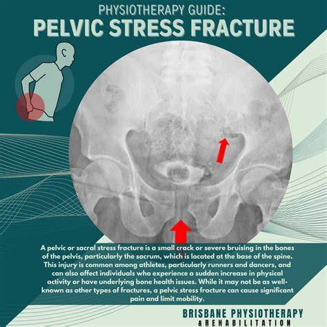
Pubic Bone Stress injury - Mr Simon Marsh
- In an athlete with groin or pubic pain during running, obtain MRI to evaluate for bone marrow edema and stress reaction at the pelvis if radiographs are negative.
- Initiate a graded return-to-run protocol aligned with pain resolution and imaging findings, ensuring the plan lasts at least as long as time off from running.
- Incorporate a progressive lower-limb and pelvic stabilizing strengthening program early in rehab to address biomechanical risk factors.
- Collaborate with pelvic physiotherapists and multidisciplinary teams to tailor rehabilitation and return-to-sport decisions.