Overview of cherry angiomas and their appearance on the hairline
Cherry angiomas are small benign growths of small blood vessels that occur in almost all patients over the age of thirty. They can be as small as only 1 - 2 millimeters in diameter, or range in size up to a centimeter. Cherry angiomas may appear flat, or may become raised over time. Sometimes a patient may have only one lesion, or patients can have dozens of them. Most of the time, patients will notice the development of these lesions on the scalp, face, trunk, and extremities. Cherry angiomas can range in color from a bright cherry red to a dark, almost purple color, commonly seen in older patients and with larger lesions. They are almost always few to no symptoms unless they have been traumatized, for instance, being cut when shaved over, etc. Once the lesions appear, they are permanent. Cherry angiomas are usually red and smooth, with even borders. They should not rapidly increase in size, scab, scale, or bleed. Lesions that exhibit these symptoms should be promptly evaluated by a dermatology provider.
Clinical characteristics and diagnosis
Angioma or haemangioma describes a benign vascular skin lesion. A cherry angioma is a small papular angioma and is a true capillary haemangioma. Cherry angiomas markedly increase in number from about the age of 40, so it has been estimated that 75% of people over 75 years of age have them. Although they are also called senile angiomas, they can occur in young people too - 5% of adolescents have been found to have them. Cherry angiomas are very common in males and females of any age or race, with no difference in sexes or races affected. They are however more noticeable in white skin than in skin of color. There may be a family history of similar lesions. The cause of cherry angiomas is unknown.
Cherry angioma is an asymptomatic firm red, blue, or purple papule, 0.1-1 cm in diameter. When thrombosed, it can appear black in color until examined with a dermatoscope when the red or purple color is more easily seen. Cherry angiomas are usually multiple. Cherry angioma is usually diagnosed clinically and investigation is not necessary for the majority of lesions. When there is uncertainty about the diagnosis, a biopsy may be performed. The angioma is composed of venules in a thickened papillary dermis. Collagen bundles may be prominent between the lobules.
Implications of location on the hairline and cosmetic considerations
Cherry angiomas on the hairline can be more noticeable due to hair growth and exposure. They are harmless and do not typically affect overall health. However, cosmetic concerns are common, leading patients to seek removal for aesthetic reasons or if the lesion is irritated by shaving or friction.
Treatment options and cosmetic management
Cherry angiomas are harmless, so they do not have to be treated. Nevertheless, there are several treatment options available for removal of these lesions; however, treatment is usually considered cosmetic. When removal is pursued, options include laser therapies and electrocautery, among others, chosen based on lesion characteristics and patient preference. The effectiveness and risks of each option should be discussed with a dermatologist. Treatments aim to reduce visibility, prevent irritation, and address patient cosmetic satisfaction. Some patients may remain satisfied with the natural appearance, especially if the lesions are stable and asymptomatic.
Prognosis and follow-up
Once the lesions appear, they are permanent. Lesions on the hairline, like other sites, typically do not spontaneously regress. If a lesion rapidly increases in size, bleeds, or changes in color or texture (such as becoming scab-like or ulcerated), urgent evaluation by a dermatologist is recommended to rule out other conditions.
Notes on research and data interpretation
Research has explored the genetic and molecular aspects of cherry angiomas, including activating mutations and the biology of capillary hemangiomas. Studies emphasize that while these lesions are common and generally benign, the exact cause remains unclear and may be multifactorial, including age, skin type, and vascular biology. Reviews on laser and light treatments highlight state-of-the-art approaches for vascular lesions, applicable to cherry angiomas when removal is desired for cosmetic reasons.
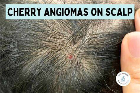
Cherry Angiomas
Practical guidance for patients
- Observe lesion stability: redness, shape, and size changes should prompt dermatologist evaluation.
- Avoid trauma to lesions, especially on the hairline where shaving may irritate them.
- Discuss cosmetic goals with a dermatologist to choose appropriate removal options if desired.
| Characteristic | Typical presentation |
|---|---|
| Size | 0.1-1 cm |
| Color | Bright cherry red to dark purple |
| Location | Scalp, face, trunk, extremities |
| Symptomatology | Usually asymptomatic unless traumatized |
| Prognosis | Permanent once appeared; generally benign |