Primary lymphoid tissues are sites of foreign antigen-independent lymphoid differentiation. B cell precursors originate, and undergo the early stages of differentiation, in the bone marrow, and enter the circulation as mature naïve B cells. The secondary lymphoid tissues include the lymph nodes, spleen, and mucosa-associated lymphoid tissue (MALT).
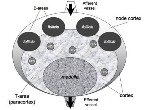
Lymphatic vessels are present throughout most of the body, and drain excess interstitial fluid from tissues, eventually returning the fluid to the circulation via the subclavian veins. Lymph nodes are small, bean-shaped structures, usually measuring between 0.2 and 2 cm, and are surrounded by a fibrous capsule. Fibrous trabeculae projecting from the capsule lend structural support to the lymph node. The lymph node can be separated into three cellular compartments: the cortex, paracortex, and medulla. The cortex contains lymphoid follicles composed mostly of B cells; in the paracortex, T cells predominate.
Lymphatic fluid containing antigens from tissues enters the lymph node via the afferent lymphatics, and flows into subcapsular, intermediate, and medullary sinuses, before exiting through the efferent lymphatics. The blood supply to the lymph node is derived from arteries that enter through the hilum of the node and branch into capillary loops that drain into postcapillary high endothelial venules in the paracortex.
The cortex of an unstimulated lymph node consists of primary follicles composed of naïve B cells, with an underlying meshwork of follicular dendritic cells. After exposure to antigen, there is a rapid proliferation of B cells. Germinal centers form in the center of B cell follicles; during this process, primary follicle cells are pushed to the periphery, where they form a mantle zone around the germinal center. The mantle zone also contains some memory B cells.
This low-power image description highlights the hilar vessels on the left, the cortex on the right with B cell follicles, and the paracortex populated by T cells and dendritic cells. Rare scattered B immunoblasts may be seen in the paracortex, large in size with vesicular chromatin and a prominent nucleolus. The medulla consists of the medullary cords, which contain lymphocytes, macrophages, and plasma cells. The medullary sinuses are contiguous with the efferent lymphatics, and contain lymph, macrophages, plasma cells, and mast cells.
The secondary follicle consists of a germinal center with a surrounding mantle zone. Uncommonly, a marginal zone composed of mature lymphocytes with moderate amounts of pale cytoplasm is seen outside the mantle zone. Germinal center B cells can be subdivided into centroblasts and centrocytes based on their morphologic features. Centroblasts are large cells with high nuclear:cytoplasmic ratios, scant basophilic cytoplasm, vesicular nuclei with smooth nuclear contours, and 1-3 small peripheral nucleoli. Centroblasts are concentrated in the dark zone of the germinal center. The centroblasts downregulate expression of surface immunoglobulin, and undergo rapid division and somatic hypermutation of their immunoglobulin genes. They then differentiate into centrocytes, express surface immunoglobulin, and migrate to the light zone, where they are selected for based on their affinity for antigens displayed on follicular dendritic cells. Centrocytes with the appropriate antigen affinity are selected to become memory B cells or plasma cells based on their interactions with dendritic cells and T cells in the light zone. Centrocytes with ineffective immunoglobulin mutations receive pro-apoptotic signals and are removed.
The classical view of the germinal center reaction suggested a unidirectional progression of germinal center B cells from the dark zone to the light zone. The main cell types in the germinal center are the germinal center B cells (GCBs), which include centroblasts and centrocytes; follicular dendritic cells; follicular helper T cells; and tingible body macrophages. It is important to note that the germinal center reaction involves very complex, and still incompletely understood, interactions between a variety of cell types. Germinal center B cells express germinal center-associated markers, including CD10 and BCL-6. CD10 is also known as CALLA, or common ALL antigen; it is expressed early in B cell differentiation, but lost as B cells mature. BCL-6 is a transcription factor that plays multiple roles in the germinal center reaction. BCL-6 suppresses p53 expression, protecting the B cell from death due to DNA damage during somatic hypermutation of the immunoglobulin genes. In a benign lymph node, CD10 and BCL-6 expression should be confined to the germinal center.
Follicular dendritic cells (FDCs) are cells of mesenchymal origin, and are more concentrated in the light zone of the germinal center. On a hematoxylin and eosin stain, follicular dendritic cells are large, often binucleate cells with vesicular chromatin and small nucleoli. FDCs express CD23 (low affinity IgE Fc receptor FcεRII), and complement receptors CR1 (CD35), CR2 (CD21), and CR3 (CD11b/CD18). FDCs display antigen in the form of antigen complexes via these surface receptors. They also secrete cytokines and chemokines that attract B cells and follicular helper T cells to the germinal center, including CXCL13, the ligand for CXCR5.
Follicular helper T cells are primarily concentrated in the light zones of germinal centers. These are specialized CD4+, CD57+, PD-1+ T cells that express BCL-6 and secrete cytokines that promote B cell proliferation and differentiation. They also play a role in selection of B cells based on affinity for antigens displayed on FDCs. Follicular helper T cells deliver survival signals to GCB cells through pathways including CD40-CD40L, PD1-PD1L, and IL-21. The pro-survival signals counteract pro-apoptotic signals from the FAS-FASL pathway. GCB cells positively selected through interactions with FDCs and follicular helper T cells become antibody-producing plasma cells or long-lived memory B cells that can differentiate into plasma cells if re-exposed to antigen. B cells not selected undergo apoptosis.
The various cell types in the germinal center can be identified by immunohistochemical stains. CD20 highlights B cells in the follicles, with both germinal center B cells and mantle zone B cells positive for CD20. In benign lymph nodes, B cells should be largely confined to follicles. The anti-apoptotic protein BCL-2 is expressed by naïve and memory B cells, and in T cells; BCL-2 is downregulated in the germinal center, making benign germinal centers negative for this marker. Immunohistochemical staining for BCL-2 helps differentiate malignant follicles of follicular lymphoma from benign reactive germinal centers. Malignant follicles of follicular lymphoma generally coexpress one or more germinal center markers with BCL-2.
In general, the lymph node architecture will be effaced in neoplastic conditions, and preserved (though possibly distorted) in reactive conditions. In reactive or infectious causes of lymphadenopathy, one or more lymph node cellular compartments may become hyperplastic. Assessment of nodal architecture is a crucial first step in evaluating a lymph node biopsy. It is important for the pathologist to have a good understanding of normal lymph node structure to recognize architectural alterations associated with lymph node disease.
Overview of Lymphoid Tissue and Spleen Context
OverviewFollicular lymphoma is a type of lymphoma. Lymphoma is cancer that affects the lymphatic system. The lymphatic system is made up of organs, glands, tubelike vessels and clusters of cells called lymph nodes. It’s part of the body's germ-fighting immune system. There are many types of lymphoma, divided broadly into Hodgkin and non-Hodgkin types. Follicular lymphoma gets its name from the fact that the cancer starts in the follicles of the lymph nodes. Lymph node follicles are groups of germ-fighting white blood cells inside each lymph node. Follicular lymphoma tends to grow slowly and is often not found until it affects many parts of the body.
CausesIt’s not clear what causes follicular lymphoma. This cancer starts inside the follicle of a lymph node. A follicle is a group of cells inside the lymph node. The follicle contains germ-fighting B cells. In infection, the follicle forms to train B cells; during this process, B cells go through changes that make them better at fighting the infection and to produce more B cells. Follicular lymphoma happens when something goes wrong during this training process, turning B cells into cancer cells that accumulate in the lymph nodes. Older age and family history are risk factors. Sometimes a follicular lymphoma can transform into a fast-growing lymphoma, such as diffuse large B-cell lymphoma.
Complications of treatment may include decreased bone density, toxicity from medicines, reactivation of viruses, and tumor lysis syndrome. Lymph nodes receive lymph via afferent channels entering the periphery and draining through subcapsular sinuses, with progression to interfollicular regions and the hilum. The B cells are distributed mainly around the periphery of the lymph node in lymphoid follicles. If B cells become active, follicles develop into secondary follicles with a central germinal center and a surrounding mantle zone. The mantle zone contains B lymphocytes that have graduated from the germinal center training camp. The interfollicular region between follicles is populated mainly by T cells.
The spleen is a distinct organ with red pulp and white pulp. The white pulp resembles miniature lymph nodes, with B cell follicles and an associated T cell population, while the red pulp contains a macrophage-rich network that clears old erythrocytes and salvages iron. The spleen can be ingested with splenomegaly and, rarely, splenic rupture. It is usually not biopsied due to bleeding risk, and many splenic conditions are inferred from clinical and imaging data rather than histology.
MALT is found in various organs, notably the GI tract, where it monitors ingested antigens. The reticuloendothelial system encompasses bone marrow, lymph nodes, spleen, MALT, thymus, tonsils, and lymphatic channels, contributing extensively to the acquired immune system. Tonsils, located in the oropharynx, mirror the standard follicular-interfollicular lymphoid architecture and are covered by stratified squamous mucosa. The organization of lymphatic channels facilitates immune surveillance by filtering lymph through strategically placed lymph nodes and tissues.
Key Cellular Players and Their Roles
The afferent lymphatics deliver antigens into the subcapsular sinus, with sinusoids rejoining to form the efferent lymphatic channel. The hilum houses arteries and veins feeding the node. Germinal center reactions involve centroblasts (dark zone) and centrocytes (light zone) with follicular dendritic cells and tingible body macrophages as co-supportive elements. Germinal center B cells express CD10 and BCL-6; CD10 is downregulated as B cells mature, and BCL-6 protects against DNA damage during somatic hypermutation.
FDCs concentrate antigens via Fc and complement receptors, shaping B cell selection. TFH cells provide survival signals through CD40-CD40L, PD1-PD1L, and IL-21, guiding affinity-based selection. Positive selection yields plasma cells and memory B cells; non-selected B cells undergo apoptosis, mediated in part by BCL-2 regulation and FAS-FASL signaling dynamics.
Immunohistochemical profiling aids differentiation between benign and malignant germinal centers. CD20 marks B cells across follicles; BCL-2 is typically absent in benign germinal centers but coexpressed with germinal center markers in follicular lymphoma. The broader architecture informs interpretation: reactive hyperplasia preserves architecture, while neoplasia may efface it.
Clinical Relevance and Diagnostic Considerations
Understanding normal lymph node structure is crucial for diagnosing lymph node disease. The germinal center reaction remains a complex, partially understood interplay among germinal center B cells, FDCs, TFH cells, and macrophages. In pathology, recognizing patterns of hyperplasia versus effacement helps differentiate reactive from neoplastic processes and informs the approach to biopsy interpretation.
- The cortex houses B cell follicles; the paracortex hosts T cells; the medulla contains plasma cells and macrophages.
- Germinal centers form during antigen exposure, leading to affinity maturation and generation of memory B cells and plasma cells.
- Immunohistochemical panels (CD20, CD10, BCL-6, BCL-2) assist in distinguishing benign reactive follicles from follicular lymphoma.