Although the ear otoscope, a tool used to look in ears, has been used pretty much exclusively by doctors for almost 400 years, modern technology is now helping parents conduct ear exams themselves. Caring parents can now easily capture real-time images of their child’s ear to share with their physician for a diagnosis. But not all ear-checking devices are the same. This article will explore the device doctors use to look into ears, what doctors look for during an ear-check exam, and why owning an otoscope can be beneficial.
What is an otoscope?
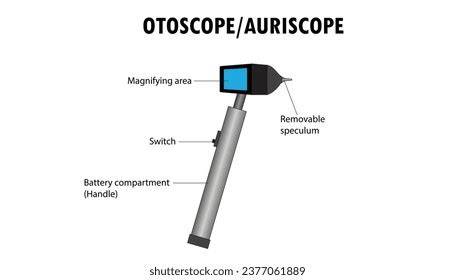
The beginning of ear-checking devices can be traced back to the 1300s when a renowned French physician and surgeon, Guy de Chauliac, envisioned a device to check ears by looking into the ears. This was the first known description of this medical tool that would one day be invented and used to check the ears of billions of people around the world. But it wasn’t until about 300 years later in the mid-1600s and late 1700s that models were developed. These early otoscopes looked more like tongs, similar to nasal speculums. The creation of the modern otoscope began in the middle of the 19th century when a German otologist named Wilhelm Kramer developed a steel speculum with a distinct funnel shape. In 1881, A. Hartmann of Berlin created an otoscope with a style that resembles what we see today. The modern otoscope, also called an auriscope, is a handheld diagnostic tool doctors use to look into the ears and ear canal. They may also use it to examine the nose and upper throat. The instrument consists of a head and a handle. The head has a magnifying lens, a light source, and a speculum. The light and magnifying lens enlarge and illuminate ear structures, allowing the clinician to visualize and evaluate the patient’s health accurately. The speculum is a viewing piece shaped like a funnel with a narrow pointed tip. Specula come in different diameters to accommodate the size of the ear passage. There are different kinds of otoscopes. A pneumatic otoscope comes with a rubber squeeze bulb that can be used to apply pressure to the eardrum to assess its movement. A video otoscope uses a small camera in the external auditory canal to visualize the ear drum. Here is what the state-of-the-art Nonagon digital otoscope, which captures high-definition video of the inner ear to share with a clinician, looks like…
What do doctors look for when using an otoscope?
Apart from the common cold, ear diseases or infections are the most common childhood illness and the most common reason parents bring their children to the doctor. In fact, five out of six children will have an ear infection before they reach three years old. Children with ear infections often tug or pull at the ear or complain of ear pain. But the only way for a physician to confirm that someone has an ear infection is for them to see what’s inside the ear and, from here, interpret the symptoms and give a diagnosis. A doctor can use an otoscope to inspect the ear canal, eardrum, and middle ear to check for an ear infection (otitis media). They will see the following during an ear examination of a healthy eardrum: eardrum appears pearly white or light grey; the ear canal, lined with some yellowish-brown ear wax; tiny bones visible in the middle ears; slight movement of eardrums when air is applied.
What does an ear infection look like through an otoscope?
In contrast, an ear with an infection (acute otitis media) may appear red, swollen, or inflamed when there is an infection. Many ear infections clear up on their own without treatment after several days. This is why many parents hesitate to go to a doctor. If that describes you, you can use an at-home otoscope like the one in the Nonagon Care Kit, which provides an easy and convenient way to examine the ear and get an accurate diagnosis without leaving your house-saving you a trip to the doctor. However, not all at-home otoscopes are the same. Some are FDA-cleared, and some are not. Some allow you to snap a photo, and some don’t. But there is only one kind (that we’re aware of) that comes in a kit, which, paired with your smartphone, can conduct nine medical exams with clinic-grade quality and share the results with your physician. Ear infections are sometimes difficult to diagnose using traditional tools, which cannot see through or around a buildup of earwax. However, impacted earwax isn’t as much of an issue when using the OtoSight Middle Ear Scope to directly visualize fluid in the middle ear. A recent clinical study conducted by PhotoniCare showed that a representative patient population had an average of ~60% occlusion of the eardrum due to earwax-for standard diagnostic tools that leaves very little of the eardrum to be examined!
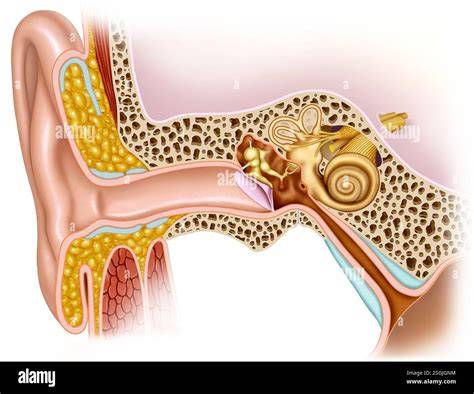
The COVID-19 pandemic has created numerous challenges for parents. An overview of the otoscope test can illustrate how a clinician views the ear canal and eardrum, sometimes aided by a binocular microscope for magnified inspection. The otoscope may have a plastic bulb on it, which delivers a tiny puff of air into the outer ear canal when pressed. This is done to see how the eardrum moves. Decreased movement of the eardrum can mean that there is fluid in the middle ear.
What is earwax and why can too much of it become an issue?
Earwax can be found in the outer ear canal. The ear canal is the tube that runs from your ear lobe to your eardrum. The skin in the ear canal has special glands that produce cerumen, which is the medical term for earwax. Once the glands produce earwax, it makes its way along the ear canal to the opening of the ear. For some people this is the end of the journey for their earwax because it falls out when the outer ear is cleaned. However, some individuals produce an above-average amount of earwax. How much earwax is produced, and what kind is produced (“wet” or “dry”) can be hereditary. For some peoples, their earwax makes its way down the ear canal and picks up dead skin cells along the way. The result is a buildup of earwax in the ear canal, which sometimes becomes hard and less mobile.
How do healthcare providers currently deal with earwax obstructions when trying to assess possible middle ear infections? Different methods for earwax removal are used depending on a patient’s complications. Oftentimes earwax can be removed with special ear drops or an in-office procedure by a healthcare provider. However, when a child or infant patient presents with impacted (clogged) earwax and is fussy or unwilling to have their ears examined, a healthcare provider is less likely to be able to accurately visualize the surface of the eardrum using an otoscope.
What can the OtoSight Middle Ear Scope do to help?
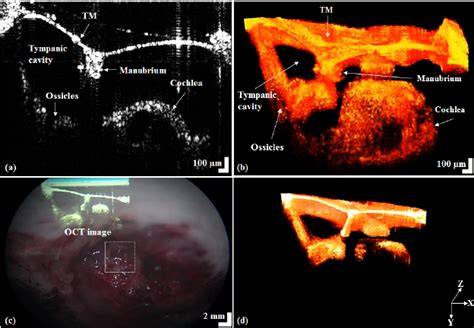
The OtoSight Middle Ear Scope is the first otoscope to provide non-invasive imaging of the eardrum and underlying middle ear. Using a novel application of Optical Coherence Tomography (OCT), OtoSight Middle Ear Scope is able to directly visualize fluid in the middle ear, a key determinant when following AAP Guidelines for diagnosing middle ear infections, particularly Acute Otitis Media (AOM). Additionally, unlike the traditional otoscope, the OtoSight Middle Ear Scope can capture images even with the presence of significant earwax build-up in the ear canal. A recent clinical study conducted by PhotoniCare showed that a representative patient population had an average of ~60% occlusion of the eardrum due to earwax-for standard diagnostic tools that leaves very little of the eardrum to be examined!
The COVID-19 pandemic has created numerous challenges for parents. Interview with Dr. PhotoniCare sits down with Dr. Overview An otoscope is an instrument which is used to look into the ear canal. The ear speculum (a cone-shaped viewing piece of the otoscope) is slowly inserted into the ear canal while looking into the otoscope. The speculum is angled slightly toward the person's nose to follow the canal. A light beam extends beyond the viewing tip of the speculum. Updated by: Ashutosh Kacker, MD, FACS, Professor of Clinical Otolaryngology, Weill Cornell Medical College, and Attending Otolaryngologist, New York-Presbyterian Hospital, New York, NY. Review provided by VeriMed Healthcare Network. Also reviewed by David C. Editorial team. The information provided herein should not be used during any medical emergency or for the diagnosis or treatment of any medical condition. A licensed medical professional should be consulted for diagnosis and treatment of any and all medical conditions. Links to other sites are provided for information only -- they do not constitute endorsements of those other sites. All content on this site including text, images, graphics, audio, video, data, metadata, and compilations is protected by copyright and other intellectual property laws. You may view the content for personal, noncommercial use. Any other use requires prior written consent from Ebix. You may not copy, reproduce, distribute, transmit, display, publish, reverse-engineer, adapt, modify, store beyond ordinary browser caching, index, mine, scrape, or create derivative works from this content. You may not use automated tools to access or extract content, including to create embeddings, vectors, datasets, or indexes for retrieval systems. Use of any content for training, fine-tuning, calibrating, testing, evaluating, or improving AI systems of any kind is prohibited without express written consent. This includes large language models, machine learning models, neural networks, generative systems, retrieval-augmented systems, and any software that ingests content to produce outputs. Any unauthorized use of the content including AI-related use is a violation of our rights and may result in legal action, damages, and statutory penalties to the fullest extent permitted by law. How the Test is Performed The provider may dim the lights in the room. A young child will be asked to lie on their back with the head turned to the side, or the child's head may rest against an adult's chest. Older children and adults may sit with the head tilted toward the shoulder opposite the ear being examined. The provider will gently pull up, back, or forward on the ear to straighten the ear canal. Then, the tip of the otoscope will be placed gently into your ear. A light beam shines through the otoscope into the ear canal. The provider will carefully move the scope in different directions to see the inside of the ear and eardrum. Sometimes, this view may be blocked by earwax. An ear specialist may use a binocular microscope to get a magnified look at the ear. The otoscope may have a plastic bulb on it, which delivers a tiny puff of air into the outer ear canal when pressed. This is done to see how the eardrum moves. Decreased movement of the eardrum can mean that there is fluid in the middle ear. How to Prepare for the Test No preparation is needed for this test. How the Test will Feel If there is an ear infection, there may be some discomfort or pain. The provider will stop the test if the pain worsens. Why the Test is Performed An ear exam may be done if you have an earache, ear infection, hearing loss, or other ear symptoms. Examining the ear also helps the provider see if treatment for an ear problem is working. Normal Results The ear canal differs in size, shape, and color from person to person. Normally, the canal is skin-colored and has small hairs. Yellowish-brown earwax may be present. The eardrum is a light-gray color or a shiny pearly-white. Light should reflect off the eardrum surface. What Abnormal Results Mean Ear infections are a common problem, especially with small children. A dull or absent light reflex from the eardrum may be a sign of a middle ear infection or fluid. The eardrum may be red and bulging if there is an infection. Amber liquid or bubbles behind the eardrum are often seen if fluid collects in the middle ear. Abnormal results may also be due to an external ear infection. You might feel pain when the outer ear is pulled or wiggled. The ear canal may be red, tender, swollen, or filled with yellowish-green pus. The test may also be done for the following conditions: Cholesteatoma External ear infection - chronic Head injury Ruptured or perforated eardrum. Risks An infection can be spread from one ear to the other if the instrument used to look inside the ear has not been cleaned well. Considerations Not all ear problems can be detected by looking through an otoscope. Other ear and hearing tests may be needed. Otoscopes sold for at-home use are lower quality than the ones used at the provider's office. Parents may not be able to recognize some of the subtle signs of an ear problem. See a provider if there are symptoms of: Severe ear pain, Hearing loss, Dizziness, Fever, Ringing in the ears, Ear discharge or bleeding.
ScopeAround Otoscope = See inside your own ear
Practical takeaways
Owning an otoscope can empower parents to monitor ear health, but professional interpretation remains important, especially when earwax obstructions or fluid behind the eardrum are involved. Some devices provide images or videos that can be shared with a clinician for a timely diagnosis. When in doubt, seek medical advice, as not all ear problems are visible with a basic exam, and additional tests may be required.
| Feature | Traditional Otoscope | Video/OCT Otoscope |
|---|---|---|
| Visualization | Limited by earwax | Can visualize fluid in middle ear |
| Imaging | Live view | High-definition imaging (OCT) |
| Accessibility | Clinic-only | At-home options available |